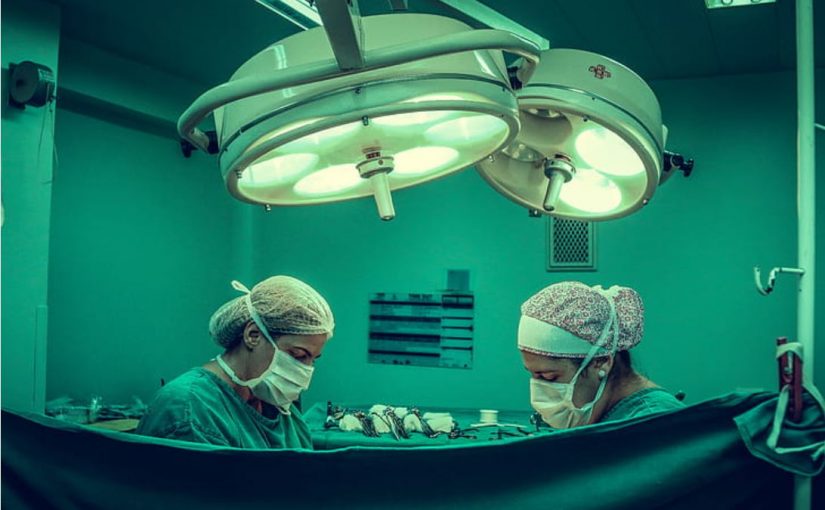
When people think about cancer surgery, they often imagine a steady-handed doctor removing a tumour from a patient’s body.
Few think about the other specialist doctor, a skilled pathologist working in the background to help the surgeon make the right decisions, often saving the patient from a second operation or futile surgery.
Using the latest equipment, operated by a highly skilled pathologist; a tissue sample is immediately frozen, sliced very thinly, stained and examined under a microscope to provide a diagnosis to the surgery team within minutes for on-the-spot action.
In cancer operations, in-theatre pathologists help surgeons determine everything from a specific diagnosis to whether they have removed all of the tumour. To the naked eye, a tumour can often look like normal body tissue. In these cases, the surgeon will cut out a small piece of tissue for the pathologist to examine.
Pathologists play an especially vital role in brain surgery. Sometimes tests before the operation cannot distinguish whether a lesion is a tumour or an infection requiring vastly different treatment. between a cancerous tumour, a benign tumour or a lesion caused by an infection.
During these operations, the surgeon will take a small sample and hand it to the pathologist who will hurry to an onsite lab to perform a mini operation on the tissue and examine it while the surgical team awaits a diagnosis. What the pathologist finds will determine whether to cut out the tumour, or to transfer the patient to a medical ward for antibiotics.
The pathologist can also tell if all the cancer has been removed, and only healthy tissue left behind; known as having negative margins.
“Without pathologists working alongside surgical teams, sometimes patients might have to go back for secondary surgery if not all the cancer had been removed,” says Dr John Ciciulla, an Anatomical Pathologist at Melbourne Pathology.
“Every time a patient goes under an anaesthetic there is a risk. We can help the surgeon perform in one surgery what may have otherwise been done in a second operation.”
“In some cases, a second operation might damage the delicate reconstructive work surgeons have performed on a patient’s face after the removal of a tumour,” he says.
“It is always better to get the tumour out on the first go and that is one of the key reasons to have a pathologist working with the surgeon.”
For other head and neck cancers pathologists are vital to ensure the tumour is correctly identified and removed. For example, tumours located in the parathyroid gland in the side of the neck. The gland is buried within the lymph nodes which can look almost identical. To a surgeon’s naked eye, it is difficult to tell the difference between the parathyroid gland and the lymph nodes, so a pathologist can examine the tissue the surgeon has removed to ensure the tumour is not left behind.
After the operation the pathologist can perform a more detailed examination of tissue inside the lab to get more information about the exact type of cancer the patient has, and this in-depth information allows treating doctors to develop the best strategy for that patient.
“There are more than 20 different subtypes of tumours in most body tissues. For example, breast cancer is not one disease. It is the pathologist who informs the oncologist exactly what they are dealing with so they can decide what treatment it will respond to,” says Dr Ciciulla.
“Each type of cancer is treated in a specific way which has an important impact on the patient’s prognosis. Although not seen by the patient, the pathologist plays a vital role in the patient outcome.”

