
Medical Scientist John Cooper set up the first pathology laboratory in the regional town of Cowra, New South Wales more than forty-five years ago and has seen enormous changes come into the lab over the years.
This predated laboratory automation and computerisation, and was a period when analyses were performed manually and all reports were hand-written. John also had daily visits from GPs who called into the laboratory during their hospital rounds to pick up pathology reports and discuss results with laboratory staff.
The Cowra lab was praised for its efficiency by Dr Harry Kramer, the Director of the Institute of Clinical Pathology and Medical Research in the 1970s.
“When I first started training in the 1960s there were no disposable plastic syringes, only glass ones that had to be washed out between patients. We also had to sharpen the needles after several uses before sterilising.
Collecting blood using a glass syringe wasn’t without its hazards as there was no auto-stop device preventing the plunger being pulled completely out of the barrel. On rare occasions, blood was spilled onto the patient’s bed making us very unpopular with the nursing staff!
We had to collect a plasma control whenever we were doing prothrombin index tests, now known as INRs. Commercial plasma controls weren’t available to us then, so we would collect additional blood from a patient who was not on anticoagulants or one of the pathology staff would volunteer.
One day we had no fresh plasma control and so I bravely volunteered, and somewhat foolishly allowed a junior medical resident to collect my blood. This proved disastrous!”
Unfortunately, the resident was not well-practised at taking blood and drew air into the syringe – John saw many bubbles inside the barrel. The resident then let go of the syringe plunger so that this air seemed to disappear into John’s arm. Air bubbles entering a vein can cause an air embolism, that can be fatal.
“The last thing I remembered was thinking ‘air embolism’ until I came to on the floor in a pool of blood, having hit my head on the central-heating unit as I collapsed. Fortunately, it was the result of a syncopal episode (dizziness), not an embolism. My head was sewn up in casualty and I resumed my day’s work!”
John remembers many changes such as when ready-made agar plates became commercially available for microbiology. Before that lab staff had to weigh out and sterilise powdered culture media to pour into plates, and horse blood was added when making blood-agar.
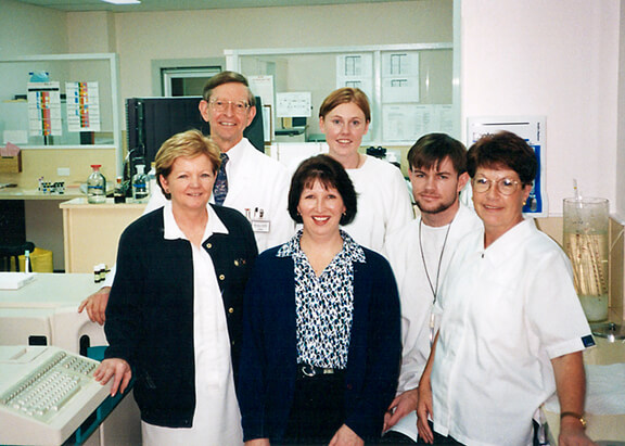
Under his stewardship, the Cowra lab grew from a single-person operation, where John was on call 24/7, to a team of 8 staff members.
One diagnosis that stands out happened during the first few weeks of John’s time at Cowra. A male patient was very ill with symptoms of meningitis and doctors were struggling to find the cause.
“An out of town referral lab had failed to detect any microorganisms in the spinal fluids submitted. The patient was getting worse, he had left hospital twice only to be readmitted.
I was sent his spinal fluid for analysis a couple of weeks after setting up the new laboratory, and using an Indian ink preparation I detected a comparatively rare form of meningitis caused by the fungus (yeast) Cryptococcus neoformans. The Indian ink preparation is a negative staining technique that microscopically reveals the large transparent capsule surrounding the yeast cell.
Torulosis, the disease caused by C.neoformans, generally starts off with a lung infection and may spread to the central nervous system. In those days it often proved fatal.
In my 35 years in pathology in was the only case of Torulosis I or the local GPs came across.”
John’s diagnosis meant the man could be appropriately treated, most likely saving his life.
